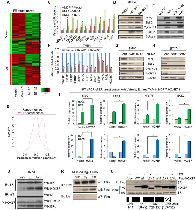
Figure 1. HOXB7 interacts with ERα and enhances expression of ER-target genes.
A, Heatmap representing relative expression of ER target genes (identified in Dutertre et al., 2010) in MCF-7-HOXB7 cells compared to MCF-7-Vector determined by microarray analysis. B, Density curves for cross-sample (n=13,182) gene expression correlations between HOXB7 and ER target genes versus randomly selected genes. Correlation between HOXB7 and ER target genes is significantly (P< 10−15) different from correlation between HOXB7 and random genes. C, Real time RT-qPCR analysis of ER target genes in stable HOXB7-overexpressing MCF-7 cells. Immunoblot analysis of HOXB7 and ER target genes in D, MCF-7-HOXB7 and E, MCF-7-TMR1 and H12 (MCF-7-EGFR/HER2) cells compared to vector control cells. F, RT-qPCR analysis of ER target genes in HOXB7 depleted MCF-7-TMR1 cells using siRNAs. Immunoblot analysis of HOXB7 and ER target genes in HOXB7-depleted G, TMR1 and H, BT474 cells compared to vector control cells. I, RT-qPCR analysis of ER target genes in MCF-7-HOXB7 stable cells incubated in estrogen deprived medium (5 % charcoal stripped serum in phenol red free DMEM) for 48 hours before treatment with vehicle, 10 nM E2, and 1 uM TAM for 24 hours. Co-immunoprecipitation (Co-IP) analysis performed J, in TMR cells using anti-ER or HOXB7 antibody and western blot analysis using anti-HOXB7 or ER antibody, or K, co-IP analysis in MCF-7-Flag-tagged-HOXB7 cells using anti-ER antibody and western blot analysis using anti-Flag antibody. L, Co-IP analysis performed with anti-flag antibodies in MCF-7 cells transfected with-Flag-tagged-full length and -deletion mutants of HOXB7 constructs. (WT: full length HOXB7, N1: N-terminal deletion 1 (1–14), N2: N-terminal deletion (38–79), WM: W129F and M130I, ΔH3: deletion of Helix domain 3 of homeodomain (183–192), ΔGlu: deletion of glutamic acid tail. Mean ± s.d. for three independent replicates. (*P<0.001).

