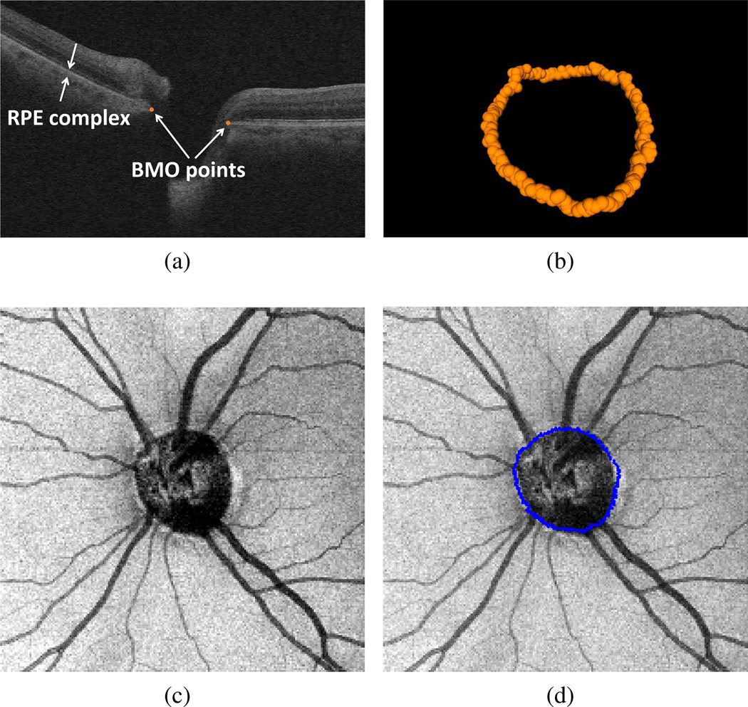
Figure 2.
Bruch’s membrane opening (BMO) within an SD-OCT volume. (a) An SD-OCT B-scan with BMO points marked with two filled circles. RPE = retinal pigment epithelium. (b) 3D view of all BMO points for the entire SD-OCT volume. (c) SD-OCT projection image. (d) Projected view of BMO points on SD-OCT projection image.

