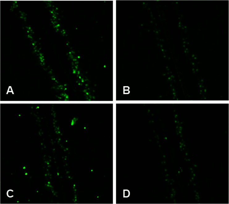
Fig. 7.
Dye transfer in A549 cells. The panels showed regions of A549 cells on slides scrape-loaded with Lucifer yellow (LY): a Control; b seawater; c seawater + STS; d PMA. Green indicated LY, including donor cells initially loaded with LY and recipient cells linked together by gap junctional intercellular communication.

