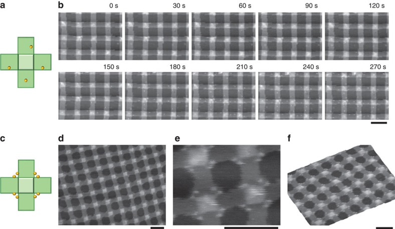
Figure 4. Surface modification of the lattice.
(a) Design of a cross-shaped DNA origami structure carrying four biotinylated staple strands (yellow dots). The top face of the origami has four biotin moieties. (b) Time-lapse AFM images of the modification of the lattice with streptavidin molecules. While scanning of the same area was ongoing, 15 μl of the folding buffer containing 20 μM streptavidin was injected into 135 μl of the standard buffer (20 mM Tris buffer (pH 7.6), 1 mM EDTA and 10 mM MgCl2), so that the final concentration of streptavidin was 2 μM. Images were obtained at a scan rate of 0.2 frames per s. The elapsed time is shown in each image. The solution of streptavidin was added at 35 s. Details are seen in Supplementary Movie 3. (c) Design of a cross-shaped DNA origami structure carrying eight biotinylated staple strands (yellow dots). (d,e) AFM images of the lattice after modification with streptavidin (2 μM). (f) A topographic AFM image of the streptavidin-modified DNA origami lattice. Scale bars, 100 nm.

