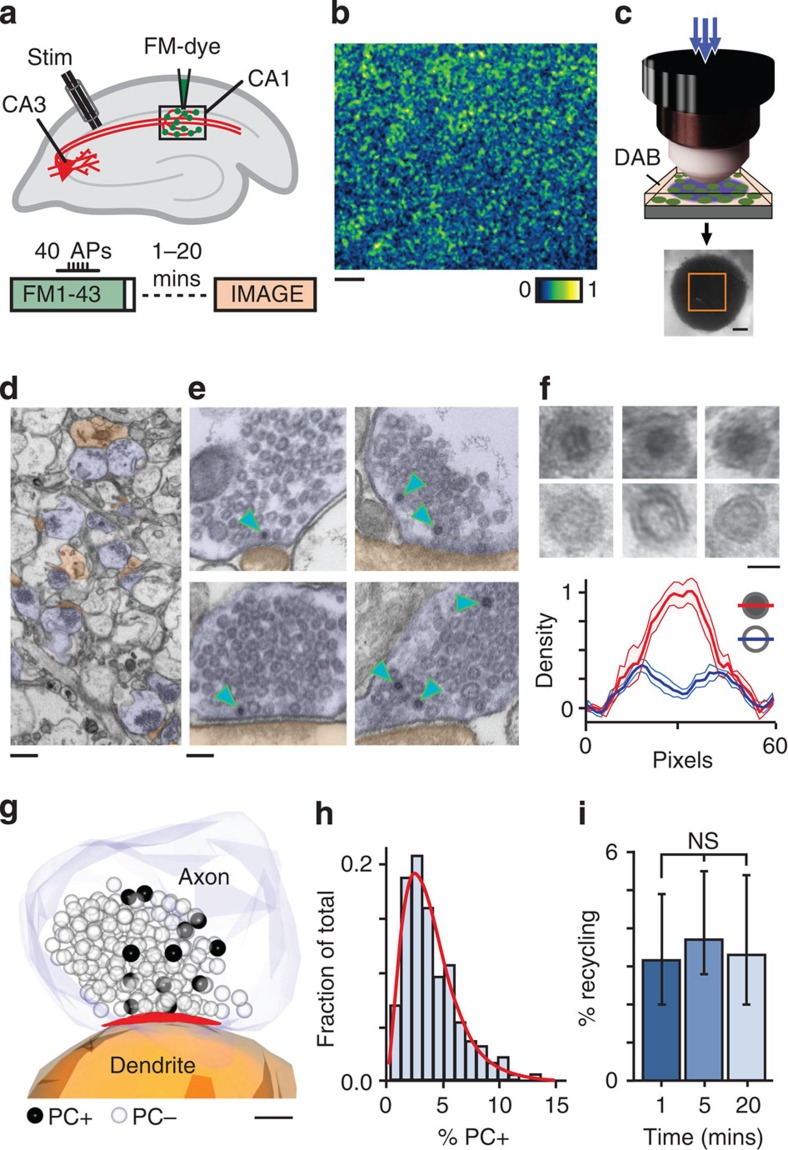
Figure 1. Ultrastructural visualization of vesicles retrieved after RRP stimulation in acute hippocampal slice.
(a) Schematic illustrating experimental protocol for labelling recycled vesicles. Extracellular stimulation (40 APs 20 Hz) of Schaffer collaterals is combined with FM-dye application to CA1. (b) Representative image of fluorescence from dye-labelled synapses in CA1. Scale bar, 10 μm. (c) Schematic illustrating approach for photoconversion of labelled vesicles. Dye-loaded fixed target region is photo-illuminated using blue light focused through an objective lens in the presence of DAB (top), producing an electron-dense spot in the slice tissue (bottom). Orange square indicates trimmed target region used for ultrastructural analysis. Scale bar, 200 μm. (d) Low magnification electron micrograph showing presynaptic terminals (blue) and postsynaptic structures (brown). Scale bar, 500 nm. (e) Typical images showing terminals with PC+ vesicles (arrowheads). Scale bar, 100 nm. (f) (Top) High magnification images of PC+ vesicles with electron-dense lumen and PC− vesicles with clear lumen. Scale bar, 25 nm. (Bottom) Cross-sectional density profiles of PC+ and PC− vesicles (n=6, mean±s.e.m.) illustrating a quantitative approach that allows for the differentiation of vesicle classes. (g) 3D reconstruction showing PC+ vesicles (dark spheres) and PC− vesicles (transparent spheres). Active zone appears red. Scale bar, 100 nm. (h) Frequency histogram of PC+ pool sizes based on ultrastructural analysis of n=187 synapses from 11 slices from 10 animals, expressed as % of total pool. Red line shows gamma function fit. (i) Summary histogram of median±IQR PC+ pool sizes for synapses fixed at different times after loading (1 min: 3.1% (2.0–4.9), n=73 PC+ vesicles from 28 synapses (including n=8 full serial reconstructions) from 3 slices from 3 animals; 5 min: 3.6% (2.5–5.6), n=138 PC+ vesicles from 59 synapses (including n=4 full serial reconstructions) from 3 slices from 3 animals; 20 min: 3.3% (2.0–5.4), n=455 PC+ vesicles from 100 synapses (including n=10 full serial reconstructions) from 5 slices from 4 animals, not significant, Kruskal–Wallis one-way ANOVA, P=0.378).

