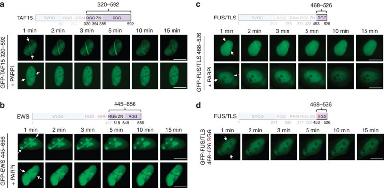
Figure 2. The RGG modules in LCD-containing proteins function as sensors of PAR formation.
(a) Recruitment kinetics of GFP–TAF15 320–592 (20 RGGs) to sites of laser microirradiation in the absence or presence of PARP inhibitor olaparib (10 μM). Time-lapse movie stills from the first 15 min after irradiation are shown. White arrows indicate the orientation of the laser line. (b) Recruitment kinetics of GFP–EWS 445–656 (16 RGGs) to sites of laser microirradiation. (c) Recruitment kinetics of GFP–FUS 468–526 (8 RGGs) to sites of laser microirradiation. (d) GFP–FUS 468–526 in which RG/RGGs were altered to SG/SGG was expressed; cells were laser microirradiated and imaged as in a–c. Scale bars, 10 μm.

