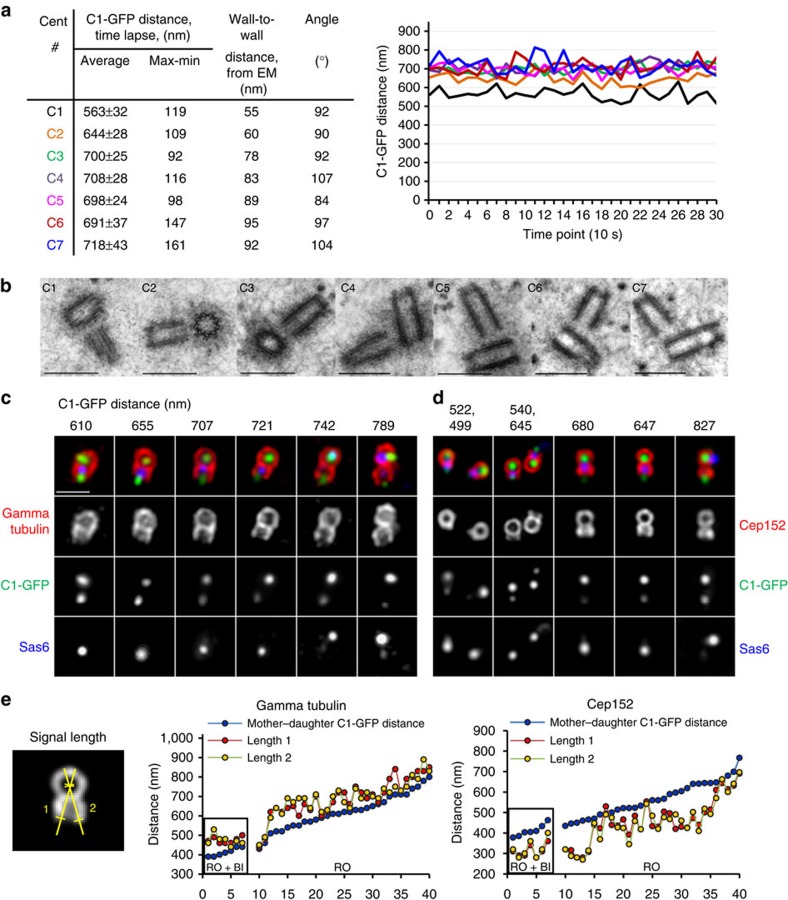
Figure 3. Analysis of ultra-structural changes within the centrosome during the earliest stages of disengagement.
(a,b) Centrioles of C1–GFP-expressing cells arrested in G2 by Cdk1 inhibitor RO-3306 (RO) were first analysed by 5 min time lapse to determine the pattern of centriole movement (expressed as the average distance between the mother and daughter C1–GFP signals). The same centrioles were then fixed and analysed by electron microscopy. Mother–daughter centriole pairs with average C1–GFP distance ≤700 nm were found in orthogonal orientation and at the wall-to-wall distance <80 nm. Daughter centrioles ≥80 nm from the wall of the mother centrioles were found disoriented. Scale bars, 500 nm. (c,d) Daughter centrioles in the centrosomes with C1–GFP distances ≤700 nm are associated with a substantial amount of PCM (as evidenced by a gamma tubulin signal) and with licensing factor Cep152. Accumulation of Cep152 precedes the degradation of Sas6, separation of two PCMs and loss of orthogonal orientation between the centrioles. Scale bar, 1 μm. (e) Quantification of daughter centriole-associated gamma tubulin and Cep152. Only centrioles with mother centriole perpendicular to the coverslip were measured. For each centrosome, two signal lengths were measured from the centre of a gamma tubulin or Cep152 toroid organized by the mother centriole, until the end of the signal associated with the daughter centriole (determined as a signal half maximum). Centrioles in cells arrested in G2 and treated with Plk1 inhibitor BI2536 (RO+BI) were measured as a control.

