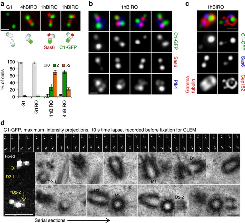
Figure 5. Plk1 inhibition in late G2 allows centriole duplication in post-mitotic cells.
(a) Analysis of centrosome-associated Sas6 signal by immunofluorescence. In control G1 cells, mother and daughter centrioles are separated and Sas6 is absent. In the 4hBIRO sample, mother and daughter centrioles are adjacent, and associated with one Sas6. 1hBIRO sample contains two adjacent centrioles associated with two Sas6 signals, indicative that mother centriole is reduplicating. Schematics of putative centriole configurations are provided to facilitate understanding. Histogram represents quantification of the cells with 0, 2 or >2 Sas6 signals. Average value and the s.d. are presented. n=300, from three independent experiments. (b) In 1hBIRO cells, one of the Sas6 signals is associated with a mother centriole (brighter C1–GFP signal) and is co-localized with Plk4 signal. (c) In centrosomes with two Sas6 signals, the older daughter centriole is organizing gamma tubulin and Cep152. Its Sas6 signal is positioned outside the Cep152 toroid organized by the mother centriole. The centre of nascent Sas6 signal co-localizes with the mother's Cep152 toroid. Scale bars, 400 nm (a–c). (d) Correlative time-lapse and electron microscopy analysis of a post-mitotic cell from 1hBIRO samples. A 5-min-long time-lapse recording with 10 s resolution reveals weak C1–GFP signals (arrows) associated with the mother centrioles, in addition to brighter C1–GFP signals. Electron microscopy further confirmed that the weak C1–GFP signals correspond to the nascent daughter centrioles (D1–2 and D2–2) positioned close to the wall of the mother centrioles. Original daughter centrioles (D1 and D2) were still in an orthogonal orientation to the mother centriole. Scale bars, 200 nm.

