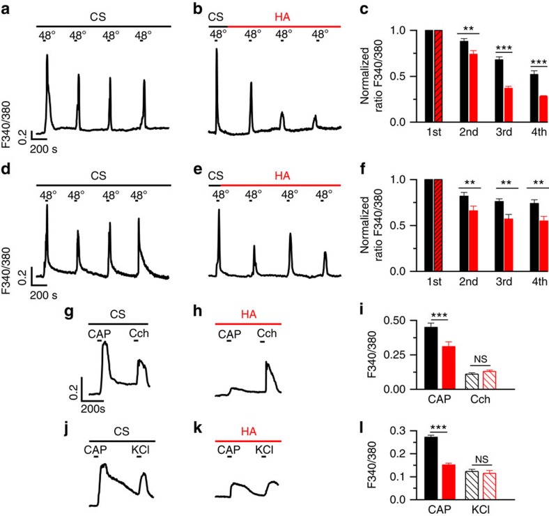
Figure 1. Inhibition by HA of intracellular calcium responses to heat (48 °C) and 100 nM CAP in HEK-TRPV1-EYFP (+) cells and DRG primary sensory neurons.
(a) Intracellular calcium rises evoked in a HEK-TRPV1-EYFP (+) cell by temperature elevations of the bathing solution to 48 °C repeated at 10 min intervals. Cytosolic Ca2+ increases are represented as the ratio of the emission fluorescence intensities at 340 and 380 nm. Notice the development of desensitization. (b) The same experiment as in a but with a HA perfusion, at the end of the first heating stimulus. (c) The normalized ratio of average amplitude change between responses evoked by successive heat pulses (indicated in the abscissae axis) in the control solution (CS, black bars) and during perfusion with HA (red bars). Striped red bar represents the average amplitude of the response in the control saline solution for cells treated with HA immediately afterwards. Notice that the inhibition was maximal at 30 min after the onset of the HA perfusion. (d–f) The same protocol as in a–c but performed in cultured adult DRG primary sensory neurons. The inhibitory effect was maximal after 20 min of HA perfusion (third versus first stimuli). (g,h) Intracellular calcium change in a HEK-TRPV1-EYFP (+) cell in response to 100 nM CAP and to 100 μM carbachol (Cch), a compound that activates endogenous muscarinic receptors in HEK293 cells, applied in CS (g) and after exposure to HA initiated 30–60 min earlier (h). (i) The average amplitude of the response to CAP (filled bars) and Cch (striped bars) under perfusion with CS (black, n=68) and in the presence of HA (red, n=100). (j–k) Intracellular calcium responses of DRG adult cultured sensory neurons to 100 nM CAP and to 30 mM KCl during perfusion with the control solution (j) and with HA (k). (l) The average amplitude of the intracellular calcium responses of DRG neurons to CAP (filled bars) or 30 mM KCl (striped bars) in control solution (black) and in the presence of HA (red). Note that the [Ca2+]i increase evoked by membrane depolarization of CAP-sensitive neurons with 30 mM KCl was not altered by HA. The data are represented as the mean±s.e.m. Student's t-test: ***P<0.001; **P<0.05; NSP>0.5.

