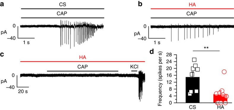
Figure 4. Reduction of the impulse response of DRG neurons to CAP during HA exposure.
Electrophysiological recordings of adult cultured DRG neurons performed in the cell-attached configuration during the application of 1 μM CAP, HP=−60 mV. (a) A sample record of the response to CAP in a single DRG neuron perfused with the control solution with a mean firing frequency of the response: 16 spikes per s (b) Sample record of CAP stimulation in a DRG neuron treated with HA and recorded under HA with a mean frequency of the response: 5 spikes per s. (c) A sample record of a DRG neuron treated and recorded in HA, in which no response to CAP was observed but an impulse discharge could be evoked with 60 mM KCl. (d) The mean firing frequency (columns) and individual data (symbols) of DRGs under CS, black (n=8), and after exposure to HA, red (n=10). Neurons that did not fire in response to KCl were excluded. Data are represented as the mean±s.e.m., Student's t-test: **P<0.01.

