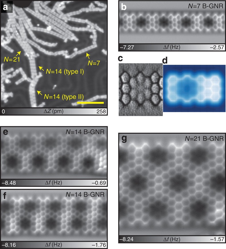
Figure 3. Fused B-GNR.
(a) STM overview of fused B-GNR. Scale bar, 10 nm. (b) Frequency shift Δf map of N=7 B-GNR and (c) the corresponding Laplace filtered image for a better view of bonds. (d) and the simulated AFM image. (e,f) Δf maps of fused N=14 B-GNR with different structures. (g) Δf map of fused N=21 B-GNR. Measurement parameters: A=38 pm and V=0 V.

