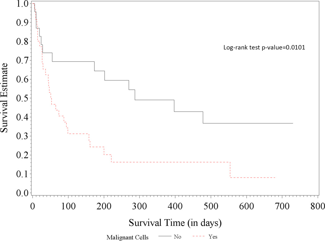
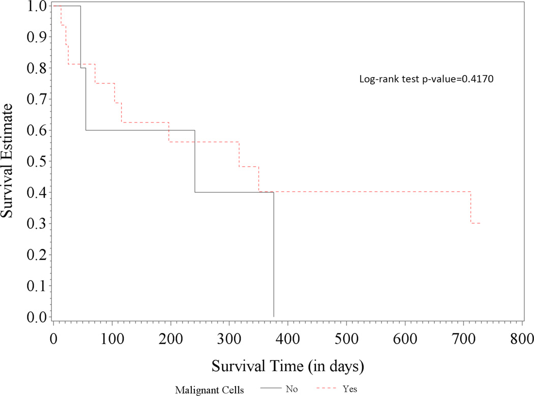
Figure 2. Access site selection for percutaneous pericardiocentesis.
Access site selection for percutaneous pericardiocentesis is based on the detection of the shortest distance to the largest fluid pocket within the pericardial space detected by echocardiography. This site selection can be achieved after careful bedside scanning from multiple directions. Needle and catheter insertion are performed while avoiding the intercostal and internal mammary arteries structures. Figure 2A represent the anterior view of the rib cage with the internal mammary arteries and intercostal arteries in red and pericardial fluid in yellow. The small green squares mark the safe access sites location. Figure 2B shows sample of short axis echocardiographic views to help select the shortest distance to the largest fluid pocket. Figure 2C shows the needle and catheter insertion site performed while avoiding the intercostal and the internal mammary arteries.


