
Fig. 3.
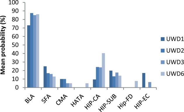
Lesion location in each UWD case quantified in terms of average probabilities for overlap with each bilateral cytoarchitectonically defined sub-region of the MTL. Although based on probability maps, these data provide strong evidence that the lesions for each UWD case were centred in the BLA and across subjects showed only minimal overlap with other MTL sub-regions if any. BL, basolateral; SF, superficial; CM, centromedial; CA, cornu ammonis; SC, subicular cortex; EC, enthorinal cortex; HA, hippocampal-amygdaloid transition area.