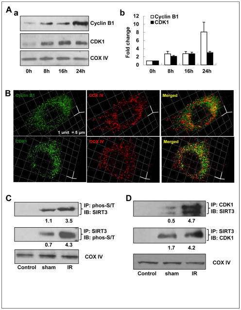
Figure 3.
SIRT3 co-localizes with CyclinB1/CDK1 in the mitochondria. A, mitochondrial accumulation of Cyclin B1 and CDK1. a, Time-course analysis of mitochondrial Cyclin B1 and CDK1 in IR-treated HCT-116 cells detected by western blot. b, Cyclin B1 and CDK1 protein levels were quantified by measuring band intensity from three western blots using Image J software and normalized with COX IV. B, representative images of mitochondrial localization of cyclin B1 (green, upper panel) and CDK1 (green, lower panel), co-stained with mitochondria marker, COX IV (red), in HCT-116 cells by 3-D structured illumination super-resolution microscopy. Scale bar, 1 unit = 5 μm. C, mitochondrial proteins were immunoprecipated (IP) with anti-phosphoserine (phos-S/T) or anti-SIRT3 followed by immunoblotting (IB) with anti-SIRT3 or anti-phos-S/T, respectively. IP with normal IgG serves as negative control and COXIV serves as equal loading control. D, co-IP of mitochondrial CDK1 and SIRT3 using mitochondrial fractions isolated from 5 Gy-irradiated or sham-irradiated HCT-116 cells (n=3).

