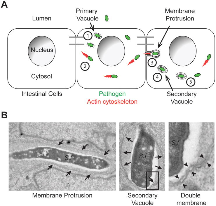
Figure 1. Sequence of events in bacterial spread from cell to cell.
(A) Cytosolic bacteria (green) spread from cell to cell within a monolayer of intestinal cells through the following sequence of events: (1) Escape from the primary vacuole, (2) Actin (red)-based motility, (3) Membrane protrusion formation into adjacent cells, (4) Resolution of membrane protrusions into (double-membrane) secondary vacuoles and (5) Escape from secondary vacuoles into the cytosol of the adjacent cell. Adapted from reference [1].
(B) Electron micrographs of the two main features of bacterial cell-to-cell spread, membrane protrusions and double membrane vacuoles. Left panel: S. flexneri (S.f) within a membrane protrusion in between two lobes of the adjacent cell nucleus (n). Membranes surrounding the protrusion are marked with arrows. Middle panel: S. flexneri within a secondary vacuole. Membranes surrounding the secondary vacuoles are marked with arrows. Right panel: high magnification showing the double membranes of a secondary vacuole corresponding to the boxed area in the middle panel. Double membranes are marked with opposing arrowheads.

