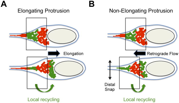
Figure 3. Dynamics of the actin cytoskeleton in protrusions.
Dynamics of the actin cytoskeleton were determined by photo-activation experiments with photo-activatable GFP-actin fusion protein. Photo-activated GFP-actin (green network) demonstrating local recycling (green arrow at the bottom) from the distal network (top panel) to the bacterial pole (bottom panel). The vertical boxes indicate the position of the actin network at the bacterial pole at the instant of photo-activation (top panel) and shortly after photo-activation (bottom panel).
(A) Cytoskeleton dynamics in elongating protrusions. The disassembly of the distal network (top panel, photo-activated actin-GFP, green) fuels the assembly of the proximal network at the bacterial pole (bottom panel, photo-activated actin-GFP, green). Note that the network at the bacterial pole did not move with reference to the plasma membrane (vertical boxes). The assembly of the network at the bacterial pole provides the forces leading to protrusion elongation (black arrow).
(B) Cytoskeleton dynamics in non-elongating protrusions. The disassembly of the distal network (top panel, photo-activated actin-GFP, green) fuels the assembly of the proximal network at the bacterial pole (bottom panel, photo-activated actin-GFP, green). Note that the network at the bacterial pole moves backwards (retrograde flow) with reference to the plasma membrane (vertical boxes). The assembly of the network at the bacterial pole provides the forces leading to retrograde flow (black arrow) and membrane scission (distal snap).

