Figure 3.
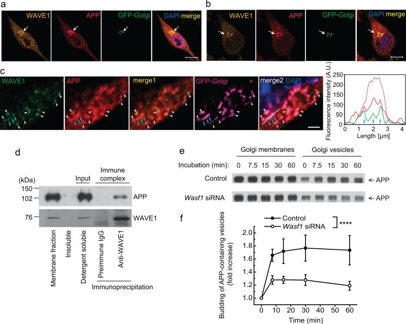
WAVE1 facilitates budding of APP-containing vesicles from the Golgi apparatus. (a–c) Immunocytochemistry of WAVE1 and APP in N2a/APPswe.PS1ΔE9 cells (a, low magnification; c, high magnification; a and c are different cells) and in N2a/APPwt cells (b). Cells were infected with a viral vector expressing a fluorescent protein fused-Golgi targeting sequence (GFP-Golgi). DAPI counterstaining was used to show the nucleus. Line scan (white line in merge 2 of c) shows coinciding fluorescence signal (cyan arrows) for WAVE1, APP and GFP-Golgi (right panel of c). Scale bars, 10 μm (a, b) and 2 μm (c). Arrows (a, b) and arrow heads (c) indicate the co-localization of WAVE1 and APP in the Golgi apparatus. (d) A detergent soluble membrane fraction from N2a/APPwt cells was used for immunoprecipitation with preimmune IgG or anti-WAVE1 antibody. WAVE1 co-precipitated with APP. (e, f) Golgi membrane (from N2a/APPwt cells) and cytosol (from N2a cells) were prepared from cells transfected with control siRNA or Wasf1 siRNA for in vitro budding assay. Reconstituted Golgi membrane and cytosol were incubated at 37 °C for the indicated times, and released vesicles were separated from Golgi membrane by centrifugation. Representative images showing time course of APP-containing vesicle formation (e) and time-dependent fold increase of APP-containing vesicle formation from six independent experiments (f, n = 6). Data represent mean ± SEM. ****P < 0.0001, Control versus Wasf1 siRNA, two-way ANOVA.

