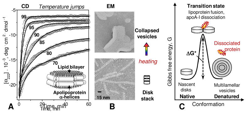
Figure 2.

Model discoidal HDL are stabilized by free energy barriers. (A) Lipoproteins reconstituted from a model phospholipid and an apolipoprotein form bilayer “disks” with protein α-helices wrapped around the perimeter. Heat denaturation kinetics of these particles was monitored in T-jump experiments from 25 °C to higher constant temperatures by CD spectroscopy at 222 nm for α-helical unfolding. Solid lines show single-exponential data fitting that was used to determine k(T) for the Arrhenius analysis. (B) Negative stain electron microscopy of discoidal HDL before and after heat denaturation. Intact particles are seen as disk stacks; such stacking is characteristic of negative staining. (C) Cartoon showing intact discoidal lipoproteins, the products of their denaturation, and the proposed high-energy transition state of disk fusion and protein dissociation.
