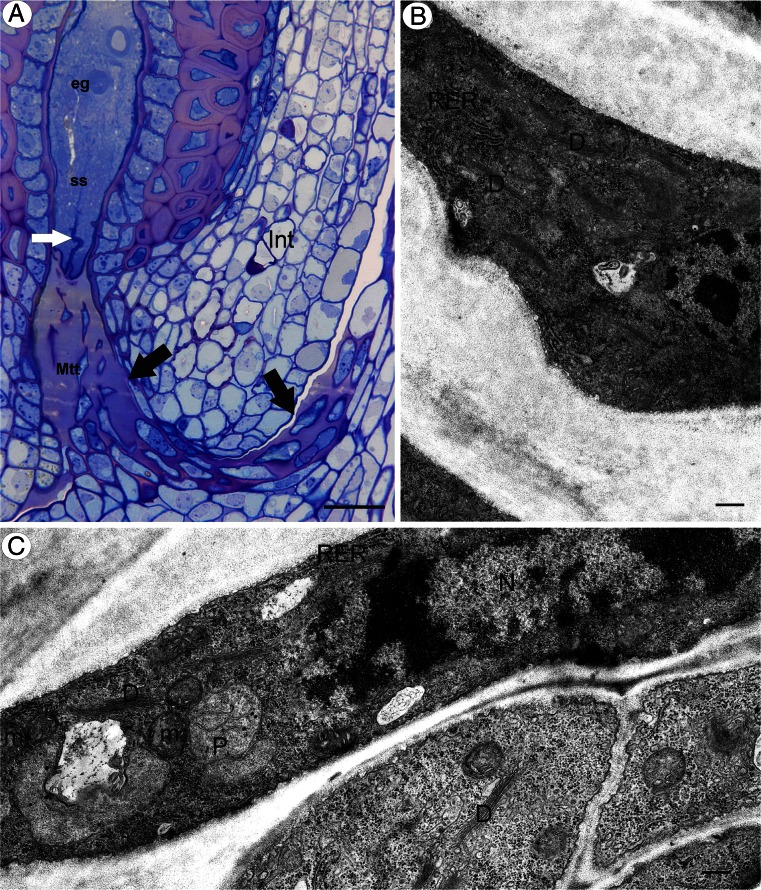
Fig. 4.
Transmitting tissue structure in apomictic dandelion T. officinale s.l. (clone SA-B). a Semithin section through an ovule and a part of an ovary showing the transmitting tissue (Mtt, black arrow), egg cell (eg), synergids (ss), and filiform apparatus (white arrow). Bar = 20 μm. b, c Ultrastructure of micropylar transmitting tissue cells, dictyosomes (D), ER cisternae (RER), plastid (P), and mitochondrion (m). Bar = 0.75 and 0.5 μm

