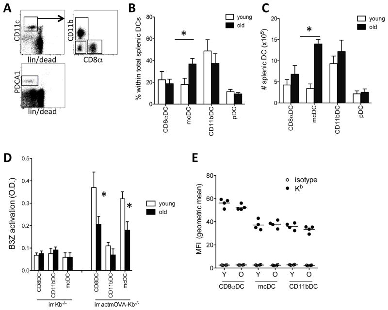
Figure 1. Altered DC composition and cross-presenting capacity in aged mice.
Spleens were harvested from young (white bars) and aged (black bars) mice and stained for CD11c, MHCII, CD11b, CD8α, in combination with a lineage stain (CD3, CD19, IgM, IgD, and NKp46) and fixable live/dead stain. A. Representative image of the gating strategy to assess DC numbers and composition. B. Frequency of DC subpopulations within the total splenic DC population. C. Absolute number of DC subpopulations per spleen. Data expressed as mean ± s.e.m. with n=9–11/group.*, p<0.05.
D. Purified young (white bars) and aged (black bars) CD8αDCs, mcDCs, and CD11bDCs were incubated with irradiated actmOVA-Kb−/− cells, resorted and cultured with the OVA257–264-specific B3Z hybridoma. B3Z activation was determined 24 hrs later by CPRG conversion assay. Data from one experiment (of 3) are expressed as mean ± s.e.m. with n=3/group.*, p<0.05. E. Fluorescent intensity if Kb on young and aged DCs. Black circles, Kb; open circles, isotype control. N=4/group.

