FIGURE 3.

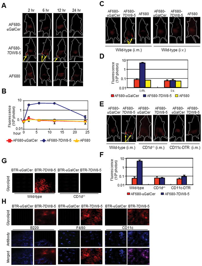
7DW8-5 is retained locally following i.m. injection and PLN-resident DCs present 7DW8-5 in a CD1d-dependent manner. (A and B) BALB/c mice (n=5/group) were injected i.m. (anterior tibialis muscles) with 5 μg AF680-αGalCer, AF680-7DW8-5 or AF680, and imaged over a time course with Lumina IVIS. (C and D) Effects of administration route on biodistribution were determined by imaging mice 8 hr after i.m. or i.v. injection of AF680-αGalCer, AF680-7DW8-5 or AF680. (E and F) CD1d−/− and CD11c-DTR mice were given AF680-labeled glycolipids by i.m. injection and imaged 8 hr later. (A, C, E) Representative images from one mouse per group are shown; yellow arrows indicate high fluorescence intensities. (B, D, F) Fluorescence intensities of the anterior tibialis muscles were quantified, and mean ± SD of five mice is shown. (G) BALB/c and CD1d−/− mice (n=5/group) were administered 2 μg BTR-αGalCer or BTR-7DW8-5 by i.m. injection. Eight-hours later, PLNs were isolated and sections were prepared for image analysis. (H) BALB/c mice (n=5/group) were treated as in (G), PLNs were isolated and stained with anti-B220, anti-F4/80 and anti-CD11c antibodies to visualize B cells, macrophages and DCs, respectively. In both (G and H), images were collected using a LSM510 confocal microscope. Images from one representative mouse are shown.
