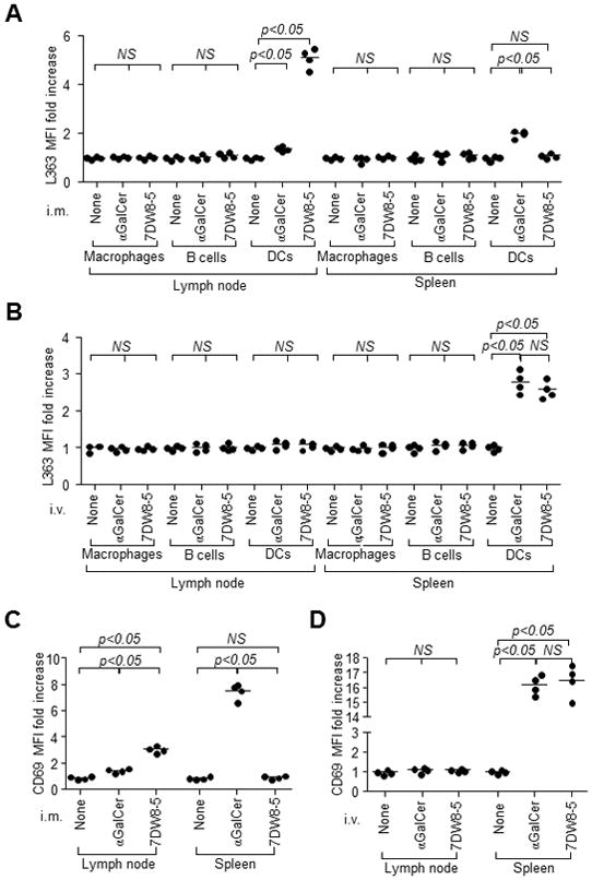
FIGURE 4.

7DW8-5 is primarily presented by PLN-resident DCs and induces iNKT cell activation in PLNs following i.m. injection. BALB/c mice (n=4/group) were administered 1 μg αGalCer or 7DW8-5 by (A) i.m. or (B) i.v. injection. PLNs and spleens were isolated 6 hr later, and CD1d-bound glycolipid on macrophages, B cells and DCs were stained with L363 and quantified by a flow cytometry. The results are expressed as fold increase of L363 MFI compared with untreated mice. BALB/c mice (n=4/group) were injected (C) i.m. or (D) i.v. with 1 μg αGalCer or 7DW8-5. Two hr later, PLNs and spleens were isolated, and iNKT cell activation was assessed by monitoring CD69 expression using flow cytometry. The CD69 MFI fold increase over mice is shown.
