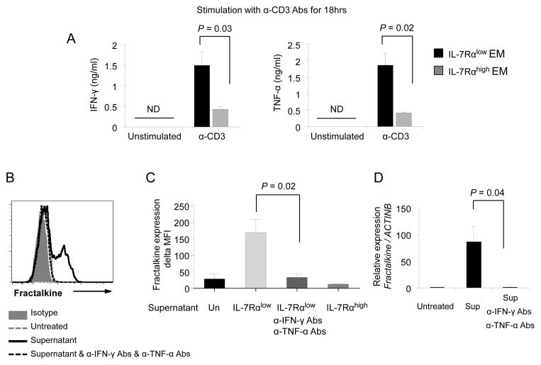
Figure 7. IL-7Rαlow effector memory CD8+ T cells produce high levels of IFN-γ and TNF-α, leading to up-regulation of fractalkine expression by HUVECs.
(A) ELISA of IFN-γ and TNF-α in culture supernatants of FACS-sorted IL-7Rαlow and high effector memory (EM) CD8+ T cells that were incubated for 18 hours with or without anti-CD3 antibodies. (B–D) HUVECs were incubated for 8 hours in the culture supernatants (Sup, 10% final concentration) of anti-CD3 antibody-stimulated IL-7Rαlow or high EM CD8+ T cells from (A) in the presence or absence of human anti-IFN-γ and TNF-α neutralizing antibodies (1μg/mL for both). Un: not treated with supernatant. (B–C) Flow cytometric analysis of fractalkine by HUVECs. (B) Representative histograms of the flow cytometric analysis. (C) Delta MFI values of fractalkine expression were obtained by subtracting MFI values of isotype control staining from MFI values of fractalkine staining. (D) RT-qPCR analysis of fractalkine gene expression in HUVECs. Bars and error bars indicate the mean and SEM, respectively (n = 3 (A), 2–5 (C–D)). ND indicates not detected.

