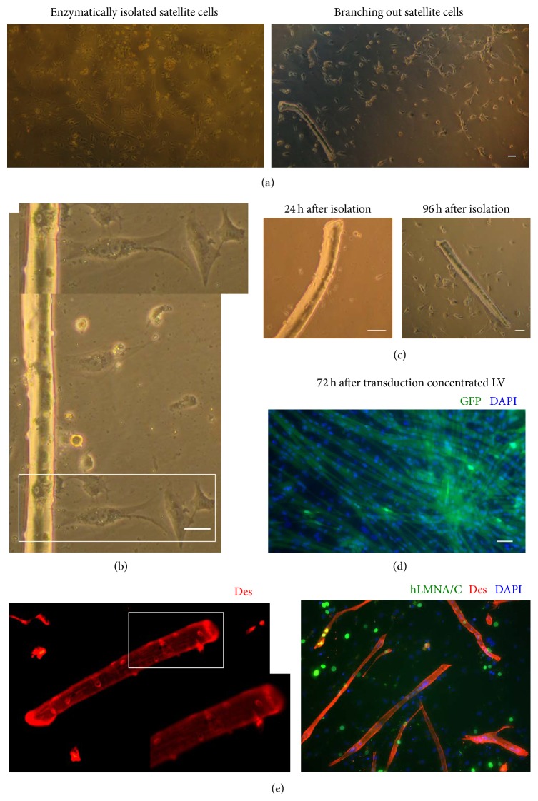
Figure 4.
Satellite cell isolation and transduction. (a) 96 hours after isolation enzymatic digestion to obtain a “pure” satellite cell culture resulted in more satellite cells in comparison to experiments where satellite cells were allowed to branch out of muscle fibers. (b) Satellite cell branching out primary muscle fiber, 96 hours after isolation. (c) Muscle fiber and branching out satellite cells 24 hours (left panel) and 96 hours (right panel) after isolation. (d) Enzymatically isolated satellite cells were transduced via concentrated LV (upper panel) encoded GFP. 72 hours after transduction via LV 95% of observed cells express GFP, thus confirming high transduction efficiency (lower panel) encoded human lamin A/C. Myotubes were stained anti-lamin and anti-desmin. Positive staining confirmed myogenicity of transduced cells. Nuclei are shown counterstained with DAPI. Scale bar corresponds to 50 μm (e) Satellite cells branching out primary muscle fiber stained anti-desmin. Positive staining confirms myogenicity of cells located on the muscle fiber surface.

