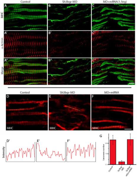
Figure 3. sh3bgr is necessary for sarcomere formation.
A-C. Stage 33 embryos were fixed and the coronal sections of somitic tissues were stained for α-actinin (Red) and MHC (Green). Knockdown of sh3bgr caused severe defects in the sarcomeres. Sh3bgr knockdown caused almost compete loss of sarcomere structures (B). Co-injection of Sh3bgr mRNA (1.5ng) with Sh3bgr-MO rescued sarcomere defects (C). D-F. Representative images of thick filaments. The fluorescent intensity plots of indicated muscle fibers are shown in (D’-F’). The average numbers of intact sarcomeres in each sample are shown in (G). Scale bar = 5μm

