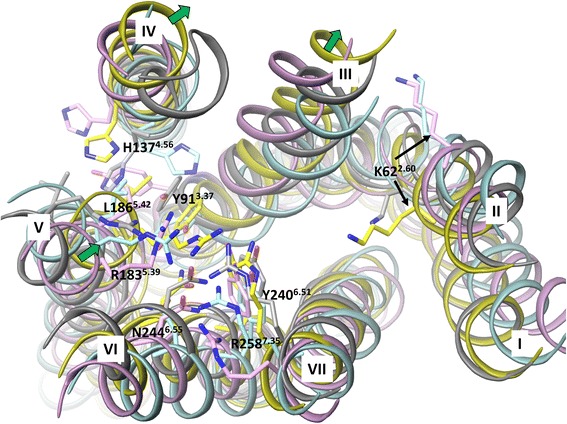
Fig. 2.

The superimposition of the FFA1 crystal structure and homology models base on the backbone of the helices in the extracellular side. The crystal structure, rhodopsin, β2 adrenergic and PAR1-based homology models are in yellow, cyan, pink and grey colour, respectively. Residues predicted to be important for ligand coordination based on mutagenesis and residue K622.60, representing the possible anchoring point for allosteric ligands are shown in stick-like representation. The green arrows indicate the large movement of helices 3, 4 and 5 in the FFA1 crystal structure compared to the homology models
