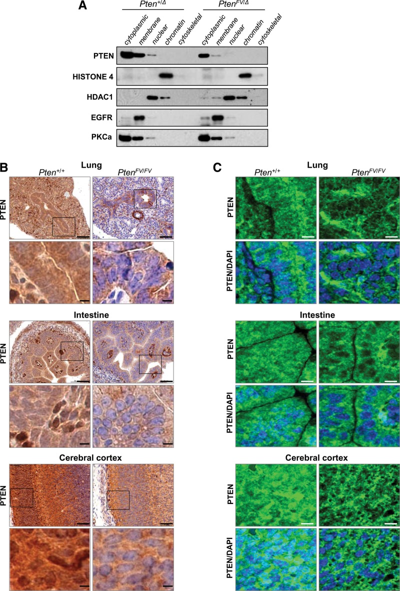
Figure 3.

PTENFV is depleted in the nucleus. (A) Cell compartment fractionation of MEFs with the indicated genotypes. Protein lysates from each fraction were immunoblotted and probed with compartment-specific markers (Histone 4, HDAC1, EGFR, and PKCa) and a PTEN-specific antibody. (B) Immunohistochemistry (IHC) of sections from the indicated organs of E15.5 Pten+/+ and PtenFV/FV embryos using a PTEN-specific antibody; IHC stained sections were counterstained with hematoxylin. The boxes in the top panels indicate regions shown at high magnification in the panels directly below. Bars: low magnification, 200 µm; high magnification, 50 µm. (C) Confocal images showing PTEN localization (green) in E15.5 embryonic tissues that were probed with the PTEN-specific antibody. Tissue sections were counterstained with DAPI (blue) to demarcate nuclei. Bar, 50 µm.
