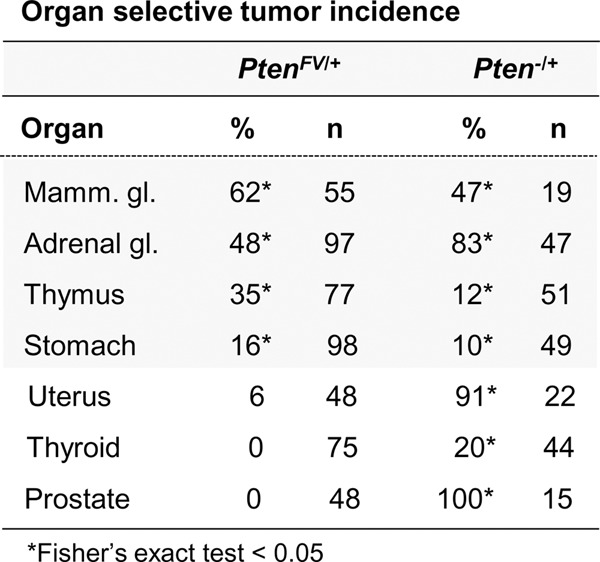
Figure 4.

Organ-selective cancer development in PtenFV/+ mice. Histopathological analysis of the indicated tissues from PtenFV/+ males and females was performed and scored as described in the Materials and Methods and Supplemental Figure 4. The table shows the percentage (%) of 18-mo-old PtenFV/+ animals with cancer in the noted organs compared with the percentage of PtenΔ/+ animals with cancer at 9 mo of age described previously (Wang et al. 2010). Carcinoma in control Pten+/+ animals was not observed. (Total n) Number of animals analyzed for each organ site. Fisher's exact tests were used to compare differences between PtenFV/+ and control Pten+/+ animals. (*) P < 0.05.
