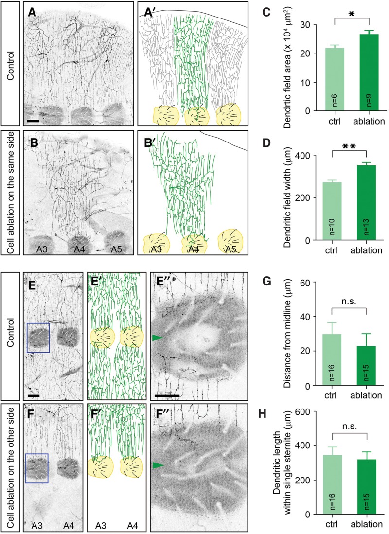
Figure 2.

Neighboring neurons are required for proper specification of the lateral boundaries but are dispensable for the ventral boundaries in v'ada neurons. (A,B) Lateral views of adult v'ada dendrites in the A3–A5 segments of an adult ventral abdomen. (A,A′) Dendrites of three v'ada neurons cover the body wall completely but redundantly. (B,B′) Ablation of neighboring v'ada neurons leads to expansion of dendritic fields of the remaining neuron. Sternites, the ventral-most epithelial region in the adult abdominal segments, are labeled in yellow. Bar, 100 µm. (C,D) Quantification of the dendritic field area (C) and the field width (D) of v'ada neurons in control and cell-ablated abdomens. Error bars indicate standard error of the mean. In C, P = 0.01. In D, P < 0.001. (E,F) Ventral views of adult v'ada dendrites in A3–A5 segments. (E,E′) Ventral boundaries of v'ada neurons are established on the periphery of sternites. (F,F′) The ventral boundaries are unaffected by ablation of v'ada neurons in the contralateral hemisegments. Magnified views of the blue boxed regions are shown in E″ and F″. Sternites are labeled in yellow. Bars: E, 100 µm; E″, 50 µm. (G,H) Quantification of the distance from the ventral midline to the branch terminals (G) and the total dendritic length within single sternites (H) in control and cell-ablated abdomens. In G, P = 0.486. In F, P = 0.669. (*) P < 0.05; (**) P < 0.01, unpaired Student's t-test.
