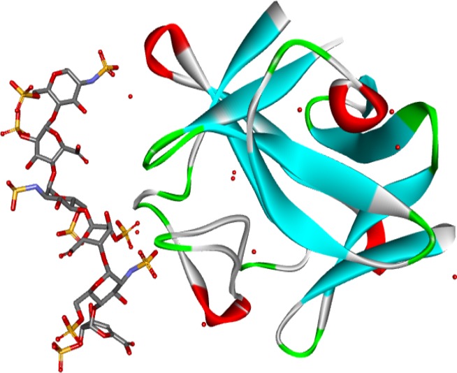
Figure 5.

Crystal structure of a heparin dp6/FGF2 complex (Faham et al. 1996). The model shows the structure of FGF2 and a heparin hexasaccharide from the pdb file 1BFC.pdb. The protein is shown as a solid ribbon coloured by secondary structure: blue for beta-strands, red for helices, green for turns and white otherwise. Water molecules are red circles. FGF, fibroblast growth factors.
