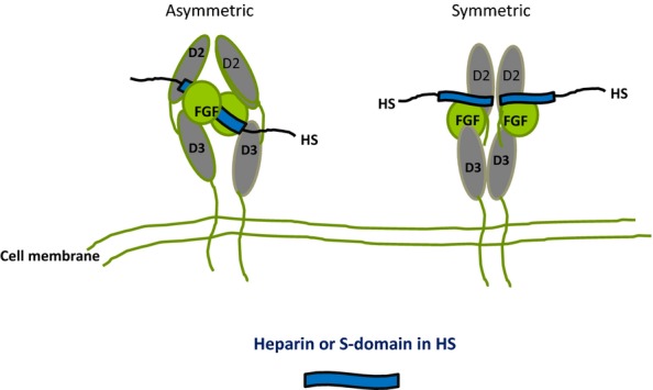
Figure 8.

Diagrammatic models of the crystal structures of FGF/FGFR/(D2 and D3 domains)/heparin complexes. In the asymmetric model (Pellegrini et al. 2000), two FGFs bind on opposite sides of a heparin dp10 saccharide and recruit two FGFRs in a stable 2:2:1 complex with minimal protein:protein contacts. In the symmetric model (Schlessinger et al. 2000), two half complexes (1:1:1 FGF:FGFR:dp10) assemble at the non-reducing ends of two dp10 heparin saccharides and these then combine, primarily by means of extensive FGFR interactions, to form the symmetric complex. In the diagrams, the heparin saccharides in the crystal structures are imagined as S domains in HS positioned internally in the asymmetric model or at the periphery of the HS chain in the symmetric version. FGF, fibroblast growth factors; HS, heparan sulphate.
