Figure 1.

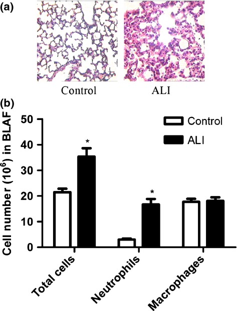
Enhanced inflammation in the lungs of mice with ALI. (a) Representative histological lung sections (HE staining) from each group. Images are shown at 250 × magnification. (b) Cell counts in BALF. LPS increases inflammatory cells in BALF in mice with ALI. Each bar represents the mean ± SEM (n = 5). *P < 0.05 compared with control group.
