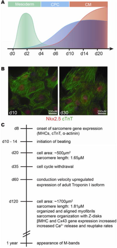
Figure 4. Molecular features of in vitro differentiated cardiomyocytes.

A) Schematic representation of gene expression patterns during the first 20 days of directed CM differentiation demonstrate temporal conservation with patterning events in mouse embryonic development. Mesodermal patterning genes (such as Mesp1 and T) are induced early and peak at day 2 (green). Markers of cardiac progenitor cells (such as Nkx2-5 and Islet1) are expressed beginning between day 4 and 6 of differentiation and are maintained in differentiated CMs (blue). Sarcomeric genes (such as aMHC and cTnT) expressed in differentiated CMs beginning between days 6 and 10 and continue to increase in expression with longer time in culture (red). B) Images of differentiatied CMs at day 10 and day 30 in culture show coexpression of Nkx2-5 (red) and cardiac Troponin T (green). C) Timeline of in vitro differentiation indicating when certain characteristics of mature CMs are acquired. Beating CMs are observed between day 10 -15 and continue to proliferate until about day 35 (88). These day 35 cardiomyocytes are still immature regarding their size, contractility, sarcomeric and mitochondrial structure (90; 92).
