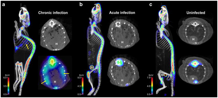
Fig. 1.

Transverse, coronal, and sagittal sections from Na[18F]F PET/CT imaging performed on a chronically M. tuberculosis-infected, b acutely M. tuberculosis-infected, and c uninfected mice, 40 min post tracer injection. Radio-densities demonstrating TB lesions are clearly visible in the lungs of infected mice (a and b), but Na[18F]F PET signal is only noted in the chronically infected mice (arrows). Na[18F]F PET signal is also noted in bones and the urinary bladder (Bl). H heart.
