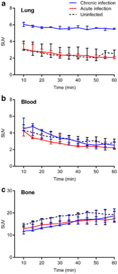
Fig. 2.

Dynamic Na[18F]F PET imaging demonstrates significantly higher signal in a pulmonary lesions from chronically infected mice (blue) compared with acutely infected (red) or uninfected (dotted black) animals. No difference is evident in b the blood or c bone compartments amongst the three different groups. Three animals were imaged for each group. Data is represented as median and interquartile range.
