
Fig. 4.
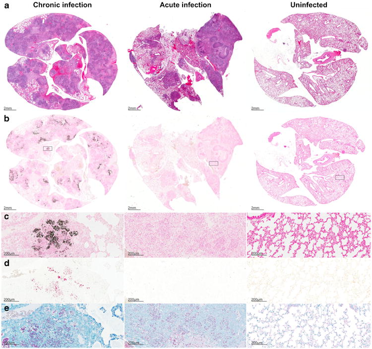
Histology sections from chronic (left panels) and acutely infected (middle panels) mice and uninfected (right panels) controls are shown. H&E staining (a), von Kossa staining for calcium phosphate deposits in black (b and c), Alizarin Red with calcium deposits visualized in red (d), and acid-fast staining for M. tuberculosis bacilli (e). Well-defined necrotic granulomas are noted on H&E staining in the M. tuberculosis-infected (acute and chronic) tissues. Von Kossa and Alizarin Red staining demonstrate deposits only in TB lesions from chronically infected tissues (b–d). AFB staining demonstrates large numbers of bacilli in infected (acute and chronic) tissues (e).