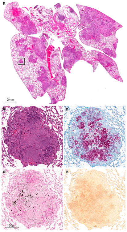
Fig. 5.

a Histology demonstrating pulmonary lesions in a M. tuberculosis-infected mouse with chronic infection. Higher magnification of a TB lesion (inset) demonstrates a classic granulomatous lesion with central necrosis (b) and a large number of M. tuberculosis bacilli seen with AFB staining (c). Microcalcifications represented as black and red deposits are evident on von Kossa (d) and Alizarin Red staining (e), respectively.
