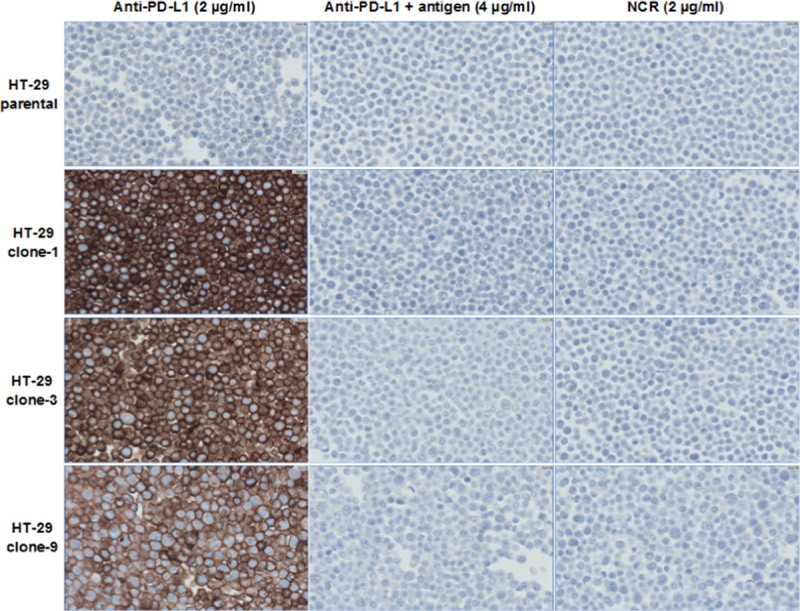
FIGURE 2.

Photomicrographs (×40) of HT-29 parental and PD-L1 overexpressing FFPE cell pellets stained by the PD-L1 IHC assay with and without antigen competition. FFPE indicates formalin-fixed paraffin-embedded; IHC, immunohistochemistry; NCR, negative control reagent; PD-L1, programmed cell death 1 ligand 1.
