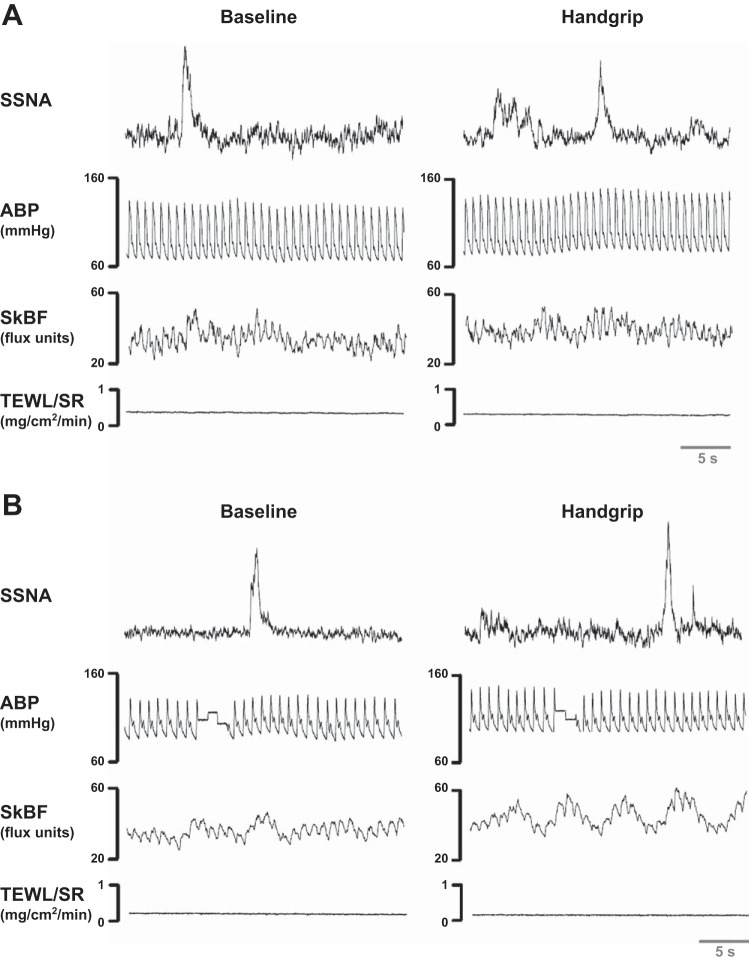
Fig. 2.
Representative neurograms during baseline and physical stress for rosacea (A) and age/sex-matched controls (B). The first row depicts the representative skin sympathetic nerve activity (SSNA) tracing during baseline (left) and during a sympathoexcitatory response to physical stress involving 2 min of 30% maximal isometric handgrip (right) in one rosacea and one control subject. The second row depicts the arterial blood pressure (ABP) waveform. The third and fourth rows depict the corresponding skin blood flow (SkBF) and transepidermal water loss/sweat rate (TEWL/SR) from cutaneous end-organs on the contralateral side of the forehead.

