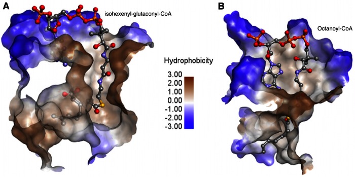
FIG 4.
Comparison of the substrate-binding mode in AtuE (A) (following docking of isohexenyl-glutaconyl-CoA onto the active site, as described in Materials and Methods) and rat ECH (B) (the PDB accession number for the experimental structure is 2DUB). Colors correspond to the hydrophobicity of the active site, as shown in the scale (blue, less hydrophobic; brown, more hydrophobic). Isohexenyl-glutaconyl-CoA and octanoyl-CoA are shown as sticks with atoms indicated by colors: gray, carbon; red, oxygen; orange, phosphorus; yellow, sulfur; blue, nitrogen.

