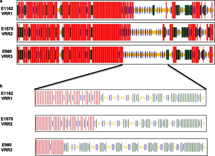
FIG 9.
Prediction of the secondary structure of SagA. Shown are comparisons of the predicted secondary structures of total SagA protein (a) and the variable repeat regions (VRRs) (b) of E. faecium strain E1162, which represents SagA protein with VRR-1; strain E1575, which represents SagA with VRR-2; and strain E980, which represents SagA with VRR-3. Secondary structures were predicted using the CFSSP (Chou & Fasman Secondary Structure Prediction Server) Web tool. Alpha-helices are indicated in red, beta-sheets in green, beta-turns in blue, and random coils in yellow.

