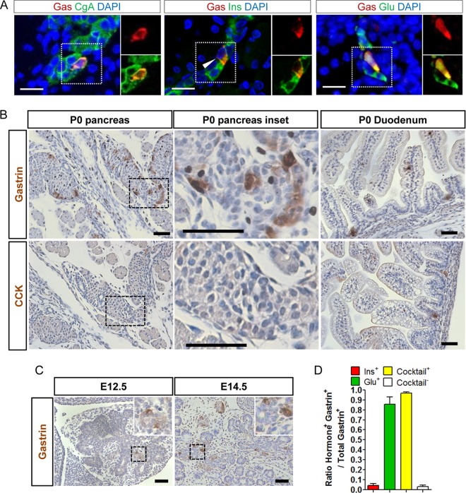
FIG 1.
Neonatal gastrin-expressing cells represent subsets of alpha and beta cells. (A) IF colocalization of gastrin and chromogranin A, insulin, or glucagon in the pancreases of newborn wild-type mice (3 ≤ n ≤ 6 mice). Magnified views of the area with the dotted outline are shown on the right. Bars = 20 μm. (B) Representative IHC stainings for gastrin and CCK in wild-type mouse pancreas and duodenum at birth. Magnified views of the area with the dashed outline are shown in the middle panel. Bars = 50 μm. (C) Representative IHC staining for gastrin in wild-type mouse pancreas at E12.5 and E14.5 (n = 3 mice of each age). (Insets) Magnified views of the area with the dashed outline. Bars = 50 μm. (D) Percentage of gastrin-expressing cells (3 ≤ n ≤ 7 mice) expressing glucagon, insulin, or any of the pancreatic hormones (Cocktail) (see Results) at birth in wild-type mouse pancreas. DAPI, 4′,6-diamidino-2-phenylindole; Gas, gastrin; Ins, insulin; Glu, glucagon.

