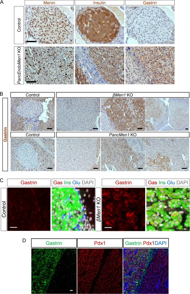
FIG 4.
PancEndoMen1 KO mice, PancMen1 KO mice, and βMen1 KO mice develop tumors expressing gastrin. (A) IHC stainings for the indicated factors in 12-month-old control and PancEndoMen1 KO mice. Note that the tumor located on the right expressed gastrin, whereas the tumor on the left did not. Bars = 50 μm. (B) IHC detection of gastrin in 12-month-old control and βMen1 KO or PancMen1 KO mice. Gastrin is not expressed in normal islet cells of control mice. Most lesions in mutant mice do not express gastrin (left), whereas subsets of tumors express gastrin at high levels (middle) or at low levels or focally (right). Bars = 50 μm. (C) Triple IF stainings for insulin, glucagon, and gastrin in 12-month-old control and βMen1 KO mice. Gastrin-expressing cells in tumors from βMen1 KO mice also expressed insulin. Bars = 20 μm. (D) Representative IF staining for Pdx1, gastrin, and DAPI (4′,6-diamidino-2-phenylindole). The lesion on the right did not express gastrin but expressed Pdx1, whereas the lesion on the left expressed both gastrin and Pdx1. Bar = 25 μm.

