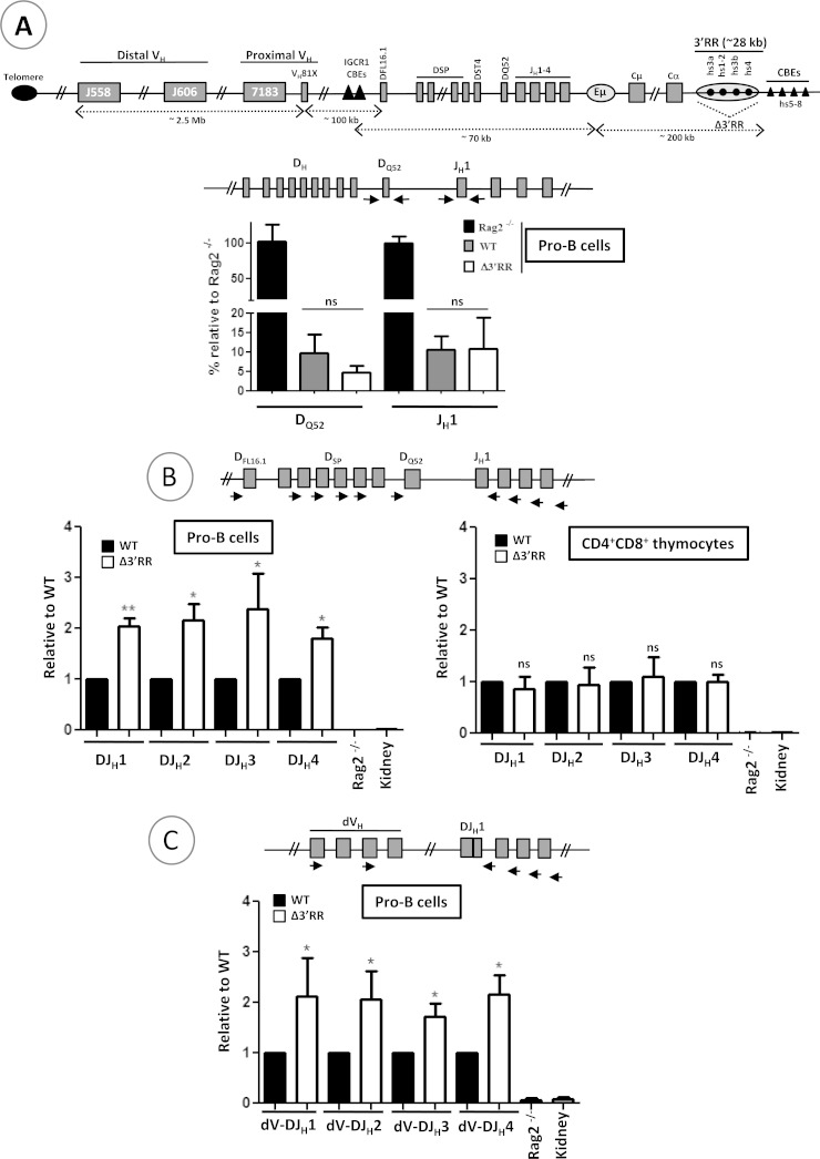
FIG 1.
V(D)J recombination in mutant mice. (A) The schematic at the top shows the mouse IgH locus (not to scale). CBEs, CTCF-binding elements. Not all CBEs are shown. Genomic DNAs were prepared from sorted WT and Δ3′RR pro-B cells and subjected to qPCR to amplify unrearranged DQ52 and JH1 gene segments. The relative positions of the primers are indicated in the schematic above the graph. Genomic DNA from Rag2−/− mice was used as a control. The hs5 sequence was used for normalization (n = 4). (B) Genomic DNAs from sorted pro-B cells and double-positive thymocytes were subjected to qPCR to quantify D-JH1, D-JH2, D-JH3, and D-JH4 recombination events. Rag2−/− and kidney DNAs were used as negative controls (n ≥ 4). (C) Quantification of distal (dVH) VH-DJH recombination events in pro-B cells by qPCR (n ≥ 4). **, P < 0.01; *, P < 0.05; ns, not significant. Error bars indicate SEM.

