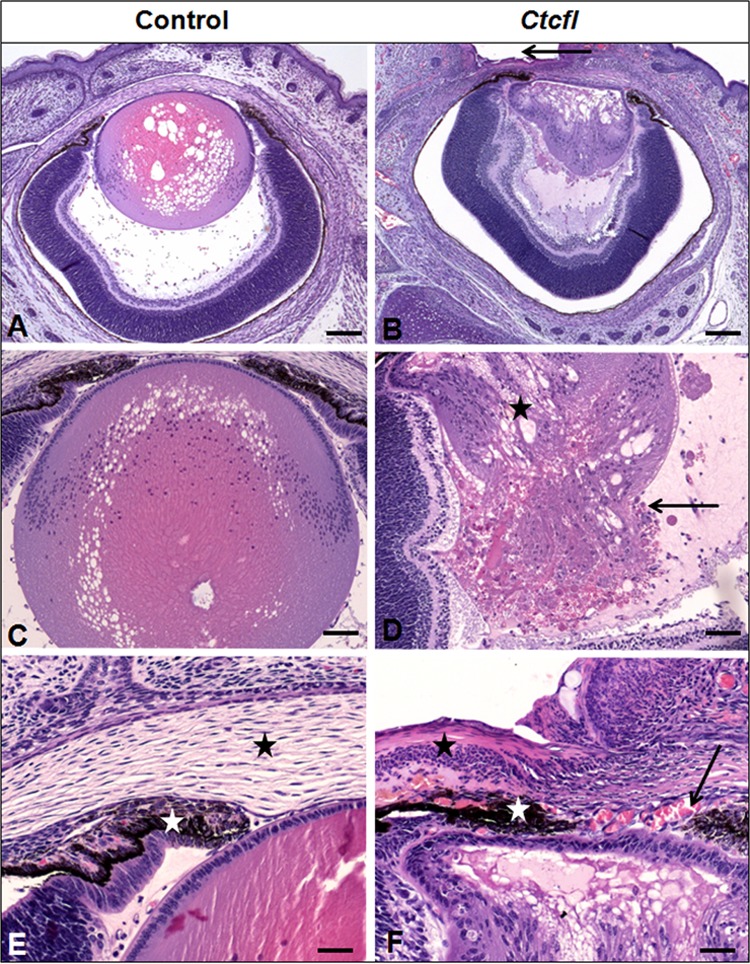
FIG 3.

Ocular lesions in Ctcfl transgenic mice. Ctcfl mice have a smaller lens (B versus A) with cataract (star in panel D) and lens rupture (arrow in panel D). Eyelids have failed to fuse (arrow in panel B), contributing to keratitis (black stars in panels F and E). The anterior lens epithelium is adhered to the interior aspect of the cornea (B, F). This is accompanied by abnormal anterior segment vascularization (arrow in panel F) and impaired iridal development (white stars in panels F and E). Control lens morphology is shown in panel C (vacuolation results from a processing artifact), and normal stromal anatomy is shown in panel E (black star). H&E staining was used. Bars: 100 μm (A, B), 50 μm (C, D), and 20 μm (E, F).
