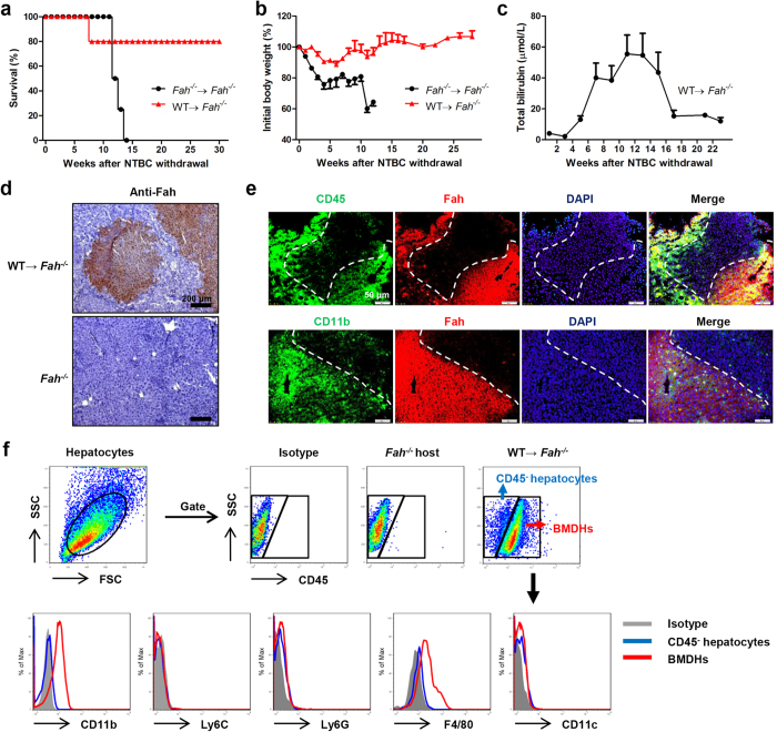
Figure 1. BMT rescues liver failure through the generation of hepatocytes in Fah−/− mice.
Survival rate (a) and body weight (b) of Fah−/− mice transplanted with Fah−/− (n = 4) or WT (n = 5) BMCs. (c) Total bilirubin levels in sera of Fah−/− mice transplanted with WT BMCs after NTBC withdrawal. (d,e) Liver tissues of WT BM-transplanted Fah−/− mice were collected 20 weeks after NTBC withdrawal and stained for Fah by immunohistochemistry (d, scale bar, 200 μm) and immunofluorescence (e, scale bar, 50 μm). The boundary of Fah+ hepatocyte area is indicated by dashed white line. (f) FACS analysis of BMDHs and Fah−/− hepatocytes 27 weeks after NTBC withdrawal. Gray shadows represent staining by the isotype control. Data are expressed as the mean ± SEM.

