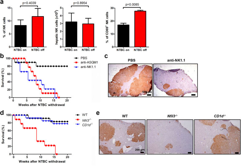
Figure 2. NK cells are essential for liver reconstitution.
(a) Hepatic NK cell (CD3−NK1.1+) percentage, absolute number, and CD69 expression in Fah−/− mice 16 weeks after WT BMT and 12 weeks after NTBC withdrawal (NTBC off); mice maintained on NTBC were used as controls (NTBC on). Fah−/− mice transplanted with WT BM were treated with anti-ASGM1 mAb (n = 9), anti-NK1.1 mAb (PK136) (n = 9), or PBS control (n = 10) throughout the period of NTBC withdrawal. (b) Survival rate is shown. (c) Liver tissues were collected from mice in the PBS-or anti-NK1.1–treated groups 14 weeks after NTBC withdrawal, and immunohistochemical staining of Fah was performed (scale bar, 500 μm). (d) Survival rate of Fah−/− mice transplanted with Nfil3−/− (n = 9), CD1d−/− (n = 14), or WT (n = 12) BM. (e) Immunohistochemical staining of Fah (scale bar, 200 μm) in liver tissues from Fah−/− mice that received BMT from the indicated mouse strain 16 weeks after NTBC withdrawal. Representative data from 2 or 3 independent experiments are shown as the mean ± SEM.

