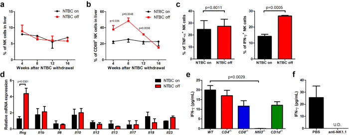
Figure 3. NK-derived IFN-γ increases after NTBC withdrawal.
Fah−/− mice were transplanted with WT BMCs, and hepatic NK cell percentage (a) and CD69 expression (b) were evaluated at the indicated time points by flow cytometry. (c)Intracellular cytokine expression in hepatic CD3–NK1.1+ NK cells was analyzed 8 weeks after NTBC withdrawal (NTBC off); mice maintained on NTBC were used as controls (NTBC on). (d) Cytokine mRNA expression in liver tissues of BM-transplanted Fah−/− mice was measured by quantitative PCR 8 weeks after NTBC withdrawal. (e) Fah−/− mice were transplanted with CD4−/−, CD8−/−, Nfil3−/−, or CD1d−/− BMCs, and serum IFN-γ levels were evaluated by ELISA 12 weeks after NTBC withdrawal. (f) Fah−/− mice transplanted with WT BM were treated with anti-NK1.1 mAb or PBS control throughout NTBC withdrawal, and serum IFN-γ levels were detected by ELISA 17 weeks after NTBC withdrawal. U.D., undetectable; Representative data from 2 or 3 independent experiments are shown as the mean ± SEM.

