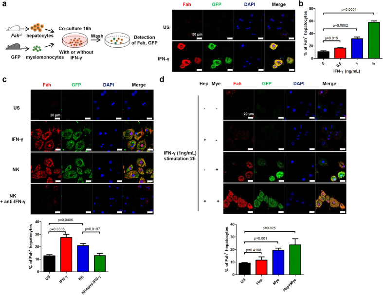
Figure 7. IFN-γ–IFN-γR interaction contributes to cellular fusion in vitro.
(a) Fah−/− hepatocytes were co-cultured with GFP+ splenic myelomonocytes for 16 h in the absence (unstimulated [US], top) or presence (bottom) of 1 ng/mL IFN-γ and then stained for Fah (red) and DAPI (blue) (scale bar, 50 μm). (b) Fah-positive hepatocytes were enumerated in the co-culture system from 5 random fields per section. (c) Fah−/− hepatocytes were co-cultured with GFP+ splenic myelomonocytes for 16 h with medium alone (US), IFN-γ (1 ng/mL), NK cells pre-stimulated with 200 IU/mL IL-2 for 24 h, or pre-stimulated NK cells plus a neutralizing anti–IFN-γ Ab (20 μg/mL); cells were then stained for Fah (red) and DAPI (blue) (scale bar, 20 μm). Representative pictures and percentage of Fah+ hepatocytes from at least 2 independent experiments are shown. (d) Fah−/− hepatocytes (Hep) or GFP+ splenic myelomonocytes (Mye) were pre-stimulated with (+) or without (−) IFN-γ (1 ng/mL) for 2 h and co-cultured together for additional 14 h. Representative images from at least 2 independent experiments for immunofluorescence staining of Fah (red) and GFP (green) and enumeration of Fah+ hepatocytes are shown(scale bar, 20 μm).

