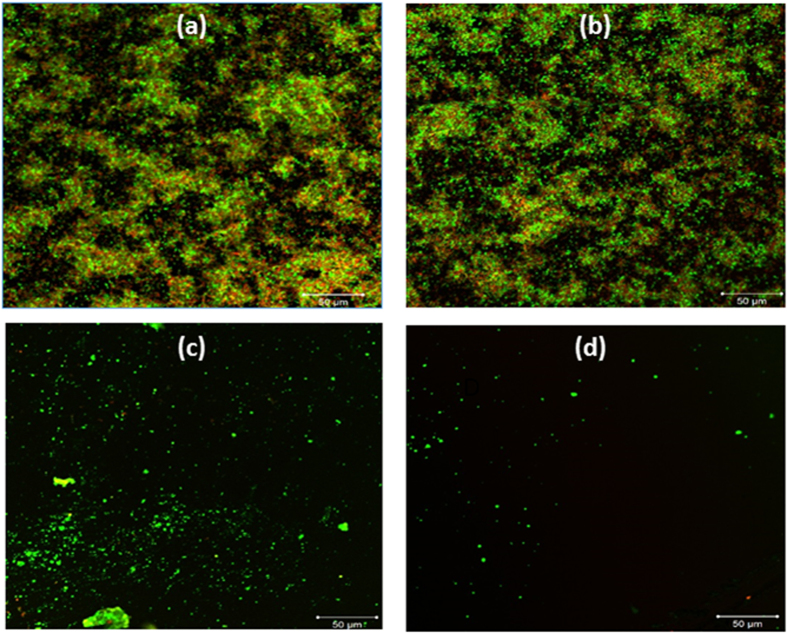
Figure 2. Confocal microscopic image of one-day grown E. coli biofilms stained with the BacLight stain (SYTO-9 and propidium iodide) after their respective treatments.
(a) Untreated control biofilm. (b) HEPES soaked biofilm. (c) E. coli biofilm after treatment with Proteinase K and DNase I. (d) E. coli biofilm treated with CO2 aerosols. The scale bars are 50 μm.

